
|
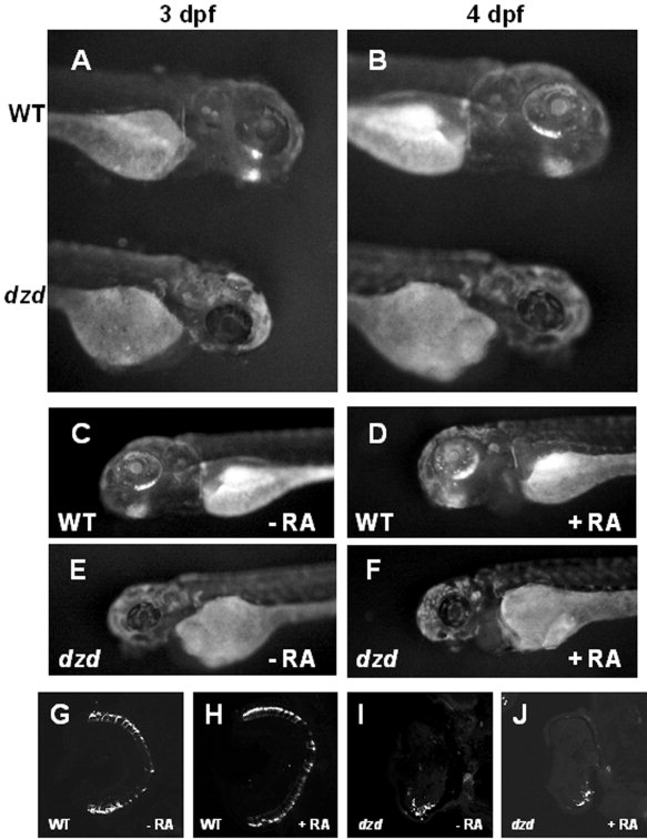
Fig. 8 Analysis of the dazed mutation in a transgenic line and after retinoic acid treatments. The dazed mutation was bred into a transgenic line (Perkins et al., [2002]) expressing a GFP-CT44 fusion protein under the control of the opsin promoter to visualize rod photoreceptors. A,B: At 3 and 4 days postfertilization (dpf), green fluorescent protein (GFP) was observed in the ventral retinas of wild-type (WT, top) but not dazed (bottom) embryos. C-F: Wild-type embryos treated from 48-96 hours postfertilization (hpf) with 300 nM retinoic acid (48-hr treatment) had more GFP expression, whereas retinoic acid had no effect on GFP expression in dazed mutants. G-J: Central, transverse sections from retinoic acid treated and control embryos were stained with 1D1 and confirmed that rods were more numerous in retinoic acid treated wild-type animals (H) than control (G) but no increase was observed in dazed mutants (I,J).