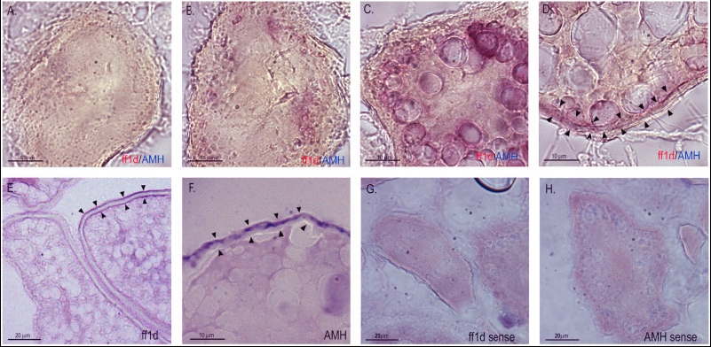
Fig. 11 Expression of ff1d and AMH in the ovary. A: Stage Ia follicle lacking AMH and ff1d expression. B: Stage Ib follicle with AMH and ff1d expression in the oocyte. C: Stage II follicle with AMH and ff1d expression in the oocyte. D: Stage III follicle with AMH and ff1d expression in cells follicular layer region. E: Stage III follicle with ff1d expression in cells follicular layer region and the inside of the vitelline envelope. F: Stage III follicle with AMH expression in cells follicular layer region. G: Stage III follicle hybridized with digoxigenin- (DIG) labeled ff1d sense probe. H: Stage III follicle hybridized with DIG-labeled AMH sense probe. Black arrowheads indicate the outer follicular layer region and the inside of the vitelline envelope. AMH, anti-Mullerian hormone.
Image
Figure Caption
Acknowledgments
This image is the copyrighted work of the attributed author or publisher, and
ZFIN has permission only to display this image to its users.
Additional permissions should be obtained from the applicable author or publisher of the image.
Full text @ Dev. Dyn.

