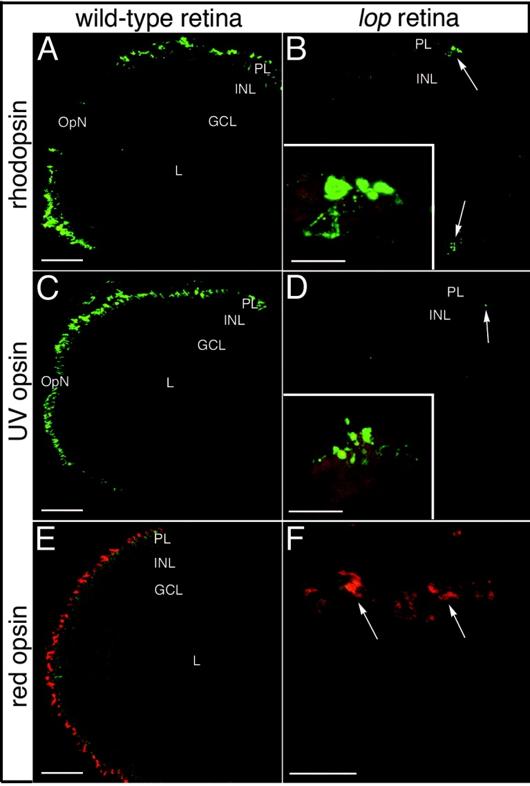
Fig. 4 Rod and cone opsin immunolocalization in wild-type and lop mutant retinas. A,C,E: Immunolocalization of rhodopsin (rods), ultraviolet (UV) opsin (short single cones), and red opsin (double cone cell member), respectively, in wild-type frozen retinal sections. B,D,F: The corresponding localization patterns in the mutant retinas. The rhodopsin and UV opsin proteins are restricted to the retinal margins of the mutants (B and D, respectively; arrows). The insets show higher magnification images of the few remaining opsin-expressing cells located at the retinal margins. Red opsin expression in the mutant retinas was also limited to the retinal marginal zone. E,F: The wild-type red opsin expression pattern and a high magnification image of the red opsin-expressing cells at the mutant retinal margin (arrows) are shown in E and F, respectively. PL, photoreceptor layer; INL, inner nuclear layer; GCL, ganglion cell layer; OpN, optic nerve; L, lens. Scale bars 50 μm in A,C,E, 20 μm in B,D,F.
Image
Figure Caption
Figure Data
Acknowledgments
This image is the copyrighted work of the attributed author or publisher, and
ZFIN has permission only to display this image to its users.
Additional permissions should be obtained from the applicable author or publisher of the image.
Full text @ Dev. Dyn.

