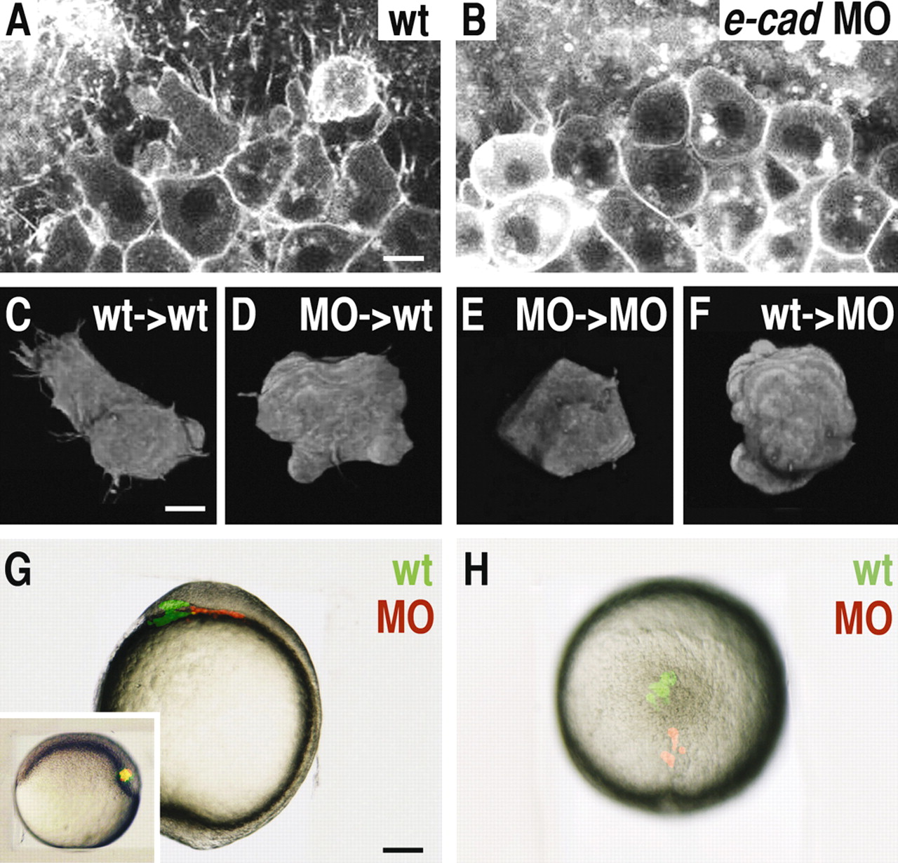
Fig. 7 Cellular morphology and position of anterior mesendodermal (prechordal plate) cells in wild-type, e-cadherin morphant and mosaic embryos at early and late stages of gastrulation. (A,B) Face- on view at the anterior edge of the prechordal plate of wild-type (A) and e-cadherin morphant (B) embryos at 6.5 hpf. (C,D) Three-dimensional reconstruction of a transplanted wild-type (C) and e-cadherin morphant (D) cell within the anterior prechordal plate of a wild-type embryo at 7 hpf. (E,F) Three-dimensional reconstruction of a transplanted e-cadherin morphant (E) and wild-type (F) cell within the prechordal plate of an e-cadherin morphant embryo at 7 hpf. (G,H) Position of transplanted wild-type (green) and e-cadherin morphant (red) cells within the prechordal plate of a wild-type embryo at 10 hpf; lateral view with anterior to the left (G), and animal view with anterior to the top (H). The inset in G shows the position of the transplanted cells (yellow) within the shield directly after the transplantation, with dorsal to the right. For all transplantation experiments (C-H), cells were transplanted at 6 hpf and the distribution (mixing) of the different cell types was monitored straight after the transplantation. MO, e-cadherin morphant cells; wt, wild-type cells. Scale bars: in A, 20 Ám; in C, 10 Ám; in G, 200 Ám.
Image
Figure Caption
Acknowledgments
This image is the copyrighted work of the attributed author or publisher, and
ZFIN has permission only to display this image to its users.
Additional permissions should be obtained from the applicable author or publisher of the image.
Full text @ Development

