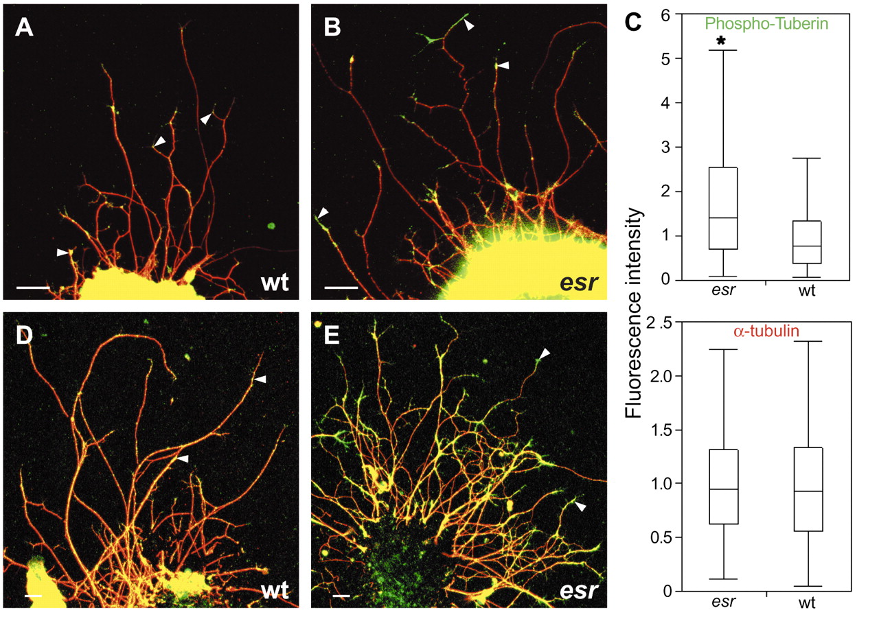
Fig. 7 Ser939 Phospho-Tuberin (green) and -tubulin (red) immunofluorescence in retinal explants. (A) After 15 hours in culture, Phospho-Tuberin (arrowheads) is detected in retinal axons, with higher levels in the esrom mutant (B). (C) At this time, Phospho-Tuberin immunofluorescence is twofold higher on average in esrom axon tips (*P<0.0001); box plot shows medians and quartiles from two normalized independent experiments (n wt=83, n esr=99 and n wt=26, n esr=78). After 26 hours in culture, the fasciculation phenotype is apparent, and Phospho-Tuberin staining difference is intensified. Scale bar: 50 Ám
Image
Figure Caption
Acknowledgments
This image is the copyrighted work of the attributed author or publisher, and
ZFIN has permission only to display this image to its users.
Additional permissions should be obtained from the applicable author or publisher of the image.
Full text @ Development

