OPTICS: MAGNIFICATION: DATE OF IMAGE: SUBMITTER COMMENTS:
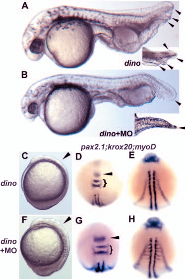
Fig. 5 Knockdown of Tsg function partially suppresses the chordino ventralization. (A) Uninjected chordino homozygote; arrowheads indicate multiple ventral fin folds, also seen in a slightly different view at higher magnification (inset). Appearance of multiple fin folds is suppressed by injection of 8 ng MO1 (B, inset shows dorsal view of tail of same embryo). (C-H) Injection of 25 ng MO5 into chordino homozygotes results in a substantial enlargement of dorsally derived tissues. Examination of live embryos at the six-somite stage showed reduced head neural tissue in C and suppression of the reduction in F (arrowheads). (C) Uninjected chordino homozygote; (F) injected chordino homozygote; lateral views, anterior is upwards. In situ analysis at the same stage with pax2.1 (arrowhead), krox20 (bracket) (D,G) and myod (E,H) showed that the somites, the MHB and rhombomeres 3 and 5 are increased in size in the injected (n=14/20) compared with uninjected mutants. (D,E) Uninjected mutants; (G,H) two views of the same injected mutant. In the injected mutant, the anterior neural and somitic mesoderm appears similar to wild type; however, the tail bud is still enlarged, indicating that the dino phenotype is not fully suppressed.
| Preparation | Image Form | View | Direction |
| not specified | still | not specified | not specified |

