
Fig. 6-S3
- ID
- ZDB-FIG-180821-54
- Publication
- Fukui et al., 2018 - Hippo signaling determines the number of venous pole cells that originate from the anterior lateral plate mesoderm in zebrafish
- Other Figures
- All Figure Page
- Back to All Figure Page
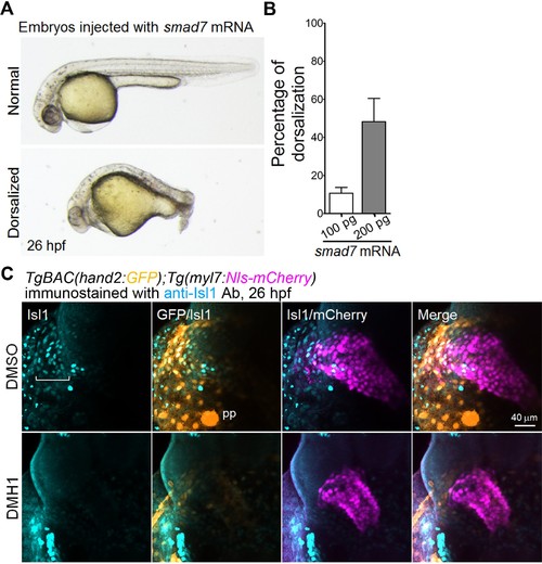
Suppression of Bmp-Smad signaling results in a decrease in the number of Isl1-positive SHF cells in the venous pole. (A) Bright-field images of embryos injected with smad7 mRNA (100 pg) at 26 hpf. The normal dorso-ventral formation (upper panel) is compared with the impaired dorso-ventral formation in the embryos injected with smad7 mRNA (lower panel). (B) Quantitative analyses of the percentage of enhanced dorsalization of the embryos injected with 100 pg or 200 pg of smad7 mRNA (n = 5). (C) Confocal 3D-stack images (at 26 hpf) of TgBAC(hand2:GFP);Tg(myl7:Nls-mCherry) embryos treated with DMSO (upper panels, n = 6) or DMH1 (10 μM, bottom panels, n = 9) from 14 hpf to 26 hpf and immunostained with the anti-Isl1 Ab. Square brackets indicate Isl1-positive cells in the venous pole. Note that both hand2- and myl7-promoter-active and Isl1-positive cells in the venous pole are absent in the embryos treated with DMH1. The confocal 3D-stack images are a set of representative images from at least three independent experiments. |