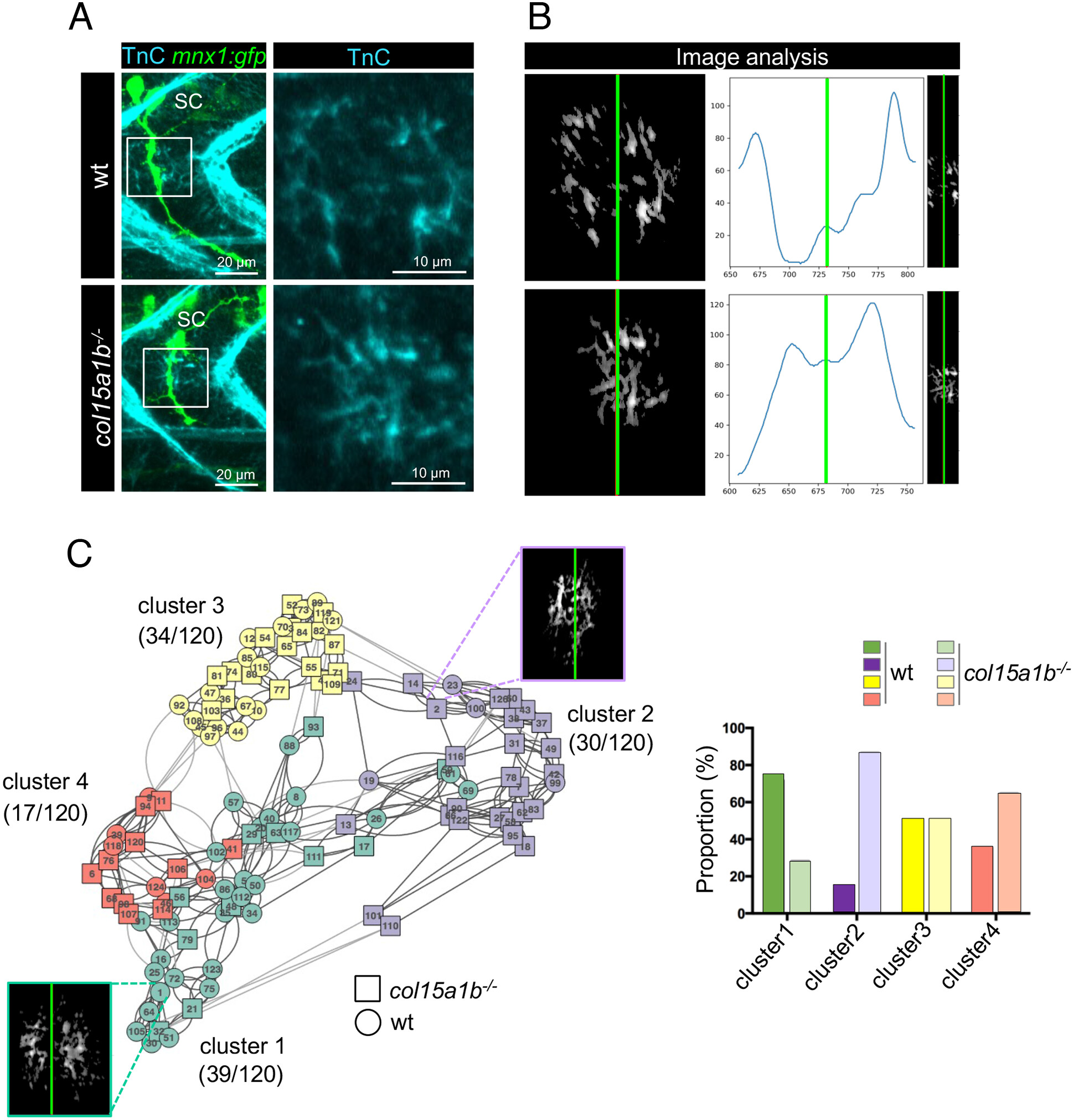
Fig. 4 Lack of ColXV-B compromises TnC channel-like organization in the motor axon path. (A) Left panel, whole-mount immunostaining of 27 hpf mnx1:gfp and col15a1b−/−;mnx1:gfp embryos with anti-TnC (cyan) and anti-GFP (green, motor axons) antibodies. Right panel, zoom of boxed images (Left) showing detail of Tnc staining. (B) Three-plot representation of one wild type and col15a1b−/−embryo. Left panel, 8-bit image resulting from the TnC deposition assessment. Right panel, 8-bit image resulting from the truncation step for TnC channel organization analysis. Curves show TnC distribution along the motor path (blue curve) estimated by vertically summing nonzero pixels then smoothed by a Gaussian filter. In all images, the green line marks the center of the TnC channel. nwt = 11 embryos, ncol15a1b−/− = 13 embryos, for each embryo 5 axons are analyzed. (C) Left panel, Clustering analysis of 120 images of TnC staining in wt (circle) and col15a1b−/− (square) mutants. One color represents one cluster and numbers in brackets indicate the total number of images for each cluster out of the 120 images analyzed. Representative eight-bit image is shown for cluster 1 (green) and cluster 2 (mauve). Right panel, Histogram showing the proportion of wt and col15a1b−/− mutant embryos in each cluster; For a same color, dark color represents wt embryos and light color represents col15a1b−/− embryos.
Image
Figure Caption
Acknowledgments
This image is the copyrighted work of the attributed author or publisher, and
ZFIN has permission only to display this image to its users.
Additional permissions should be obtained from the applicable author or publisher of the image.
Full text @ Proc. Natl. Acad. Sci. USA

