Fig. S3
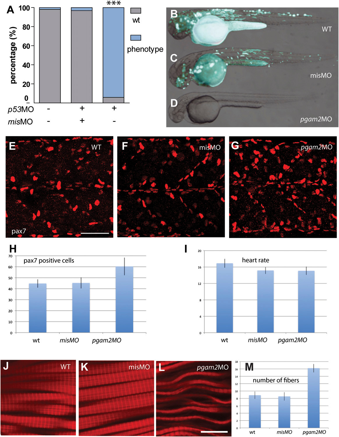
Control morpholino experiments in zebrafish. (A) Percentage of larvae showing a reduction in birefringence (phenotype, blue) after injection of the indicated morpholinos (Fisher’s exact test ; ***P < 0.0001). Injection of control morpholinos carrying five mismatches (misMO) does not lead to any obvious phenotype. (B–D) Coinjection of pgam2 morpholino and a construct carrying a GFP reporter of pgam2 expression revealed loss of GFP expression (D) compared with embryos only injected with the reporter construct (B) or to embryos coinjected with the mismatch morpholino (C). (E–H) Immunostaining against Pax7 revealed an increase of Pax7-positive nuclei in pgam2 morphants (G) compared with uninjected (F) or control morpholino-injected embryos (G). (Scale bar: 47.5 μm.) (H) Quantification of Pax7-positive myoblasts. All nuclei were counted in an area of 500 × 500 pixels in 10 different 48 hours postfertilization (hpf) embryos for each category and summarized in a chart. (I) Rate of heart beat in 48 hpf embryos (counted during 10 s, n = 12 embryos for each category). (J–M) Immunostaining with phalloidin in (J) uninjected, (K) control morpholino-injected embryos, and (L) pgam2 morphants; 48 hpf embryos. (Scale bar: 12 μm.) (M) Number of fibers present within a square of 500 × 500 pixels (eight squares counted for each category).

