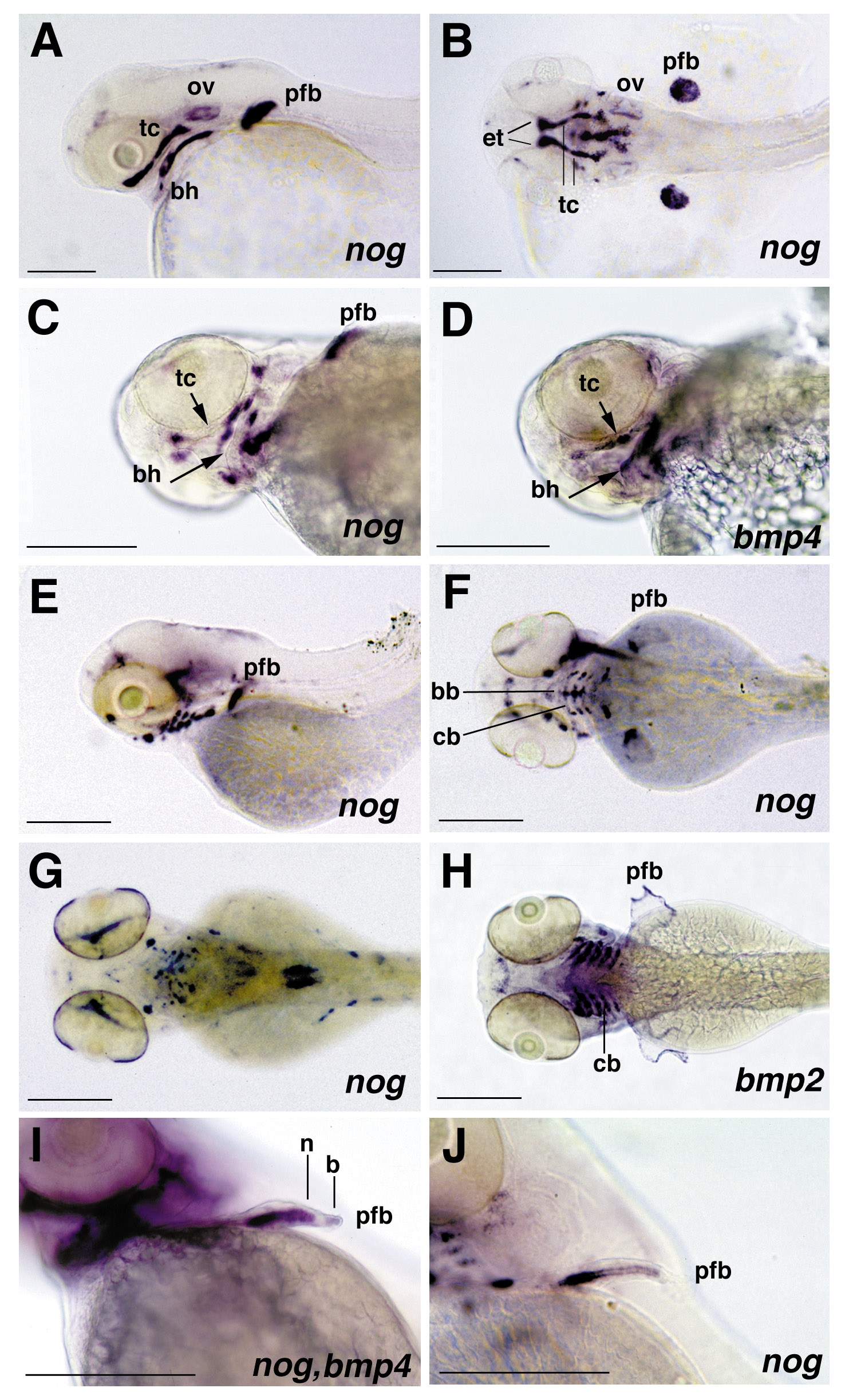
Fig. 4 Expression pattern of noggin and bmp2b and bmp4 in branchial arches and pectoral fin primordia, revealed by whole mount in situ hybridization of albino zebrafish larvae. (A and B) 48 hpf, noggin expression, lateral view (A) and dorsal view (B) on head. noggin displays a broad expression in inner regions of the pectoral fin buds (pfb), while cells at the surface lack noggin expression. In addition, noggin is strongly expressed in the ethmoid plate (et) and the trabeculae cranii (tc), components of the neurocranium, and in specific subregions of the pharyngeal arches, including the anteriormost portion of the hyoid, the basihyale (bh). Weak staining is also observed in the otic vesicles (ov). (C and D) 60 hpf, noggin expression (C) and bmp4 expression (D), ventrolateral view on head; noggin and bmp4 are expressed in complementary patterns in subregions of the trabeculae cranii and the hyoid. noggin transcripts are present in distal regions of the tc and posterior regions of the hyoid, bmp4 transcripts in central regions of the tc (indicated with short arrows) and the bh (indicated with long arrows). (E and F) 72 hpf, noggin expression, lateral view (E) and dorsal view (F); noggin is expressed in the basibranchial (bb) and ceratobranchial (cb) components of the gill arches, while no expression is detected in the pharyngeal arches. (G and H) 84 hpf, noggin (G) and bmp2b (H) expression, dorsal view; noggin expression in the gill arches has declined, while bmp2b displays strong expression in the basibranchial components of the gill arches and in apical regions of the pectoral fin primordia. (I and J) noggin (I, J) and bmp4 (I) expression in pectoral fin primordia (pfb); (I) 60 hpf, (J) 72 hpf, anterolateral view; noggin (n) is expressed in proximal regions, bmp4 (b) in distal regions of the pectoral fin primordia (I). Expression of noggin in inner cells declines in a distal-to-proximal wave. Cells with reduced noggin levels display an elongated cell shape, which is characteristic for chondrocytes (J). Abbreviations: bb, basibranchial; bh, basihyale; cb, ceratobranchial; et, ethmoid plate; ov, otic vesicle; pfb, pectoral fin bud; tc, trabeculae cranii; hpf, hours after fertilization.
Reprinted from Developmental Biology, 204, Bauer, H., Meier, A., Hild, M., Stachel, S., Economides, A., Hazelett, D., Harland, R.M., and Hammerschmidt, M., Follistatin and Noggin are excluded from the zebrafish organizer, 488-507, Copyright (1998) with permission from Elsevier. Full text @ Dev. Biol.

