Fig. 5
- ID
- ZDB-FIG-160324-18
- Publication
- Chang et al., 2016 - CDX4 and retinoic acid interact to position the hindbrain-spinal cord transition
- Other Figures
- All Figure Page
- Back to All Figure Page
|
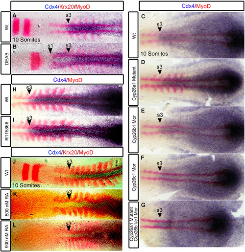
Tissue specific regulation of cdx4 expression by RA signaling. In wildtypes, cdx4 anterior expression limit corresponds to the beginning of the spinal cord located at the level of somite 3 (A, C, H, J, arrowheads). cdx4 expression is rostrally expanded to the level of somite 1 (B arrowhead) in RA-deficient embryos. Expression of cdx4 is unchanged in R115866 treated embryos (I arrowhead), in embryos treated with 500 nM RA (K arrowhead) and 900 nM RA (L arrowhead). cdx4 expression remains unchanged in cyp26a1 mutants (D), cyp26b1 morphants (E), cyp26c1 morphants (F), and cyp26a1 mutants injected with cyp26b1 and cyp26c1 morpholinos (G). (A-G, J-L) Embryos are co-stained with myoD (somites) and krx20 (r3,r5). (H-J) Embryos co-stained with myoD. (A-L) Embryos are at 10 somite stage and flat-mounted. (A-B, H-I) n=30/30, (C-G) n>12, (J-L) n=20/20 per condition. |
Reprinted from Developmental Biology, 410(2), Chang, J., Skromne, I., Ho, R.K., CDX4 and retinoic acid interact to position the hindbrain-spinal cord transition, 178-89, Copyright (2016) with permission from Elsevier. Full text @ Dev. Biol.

