
|
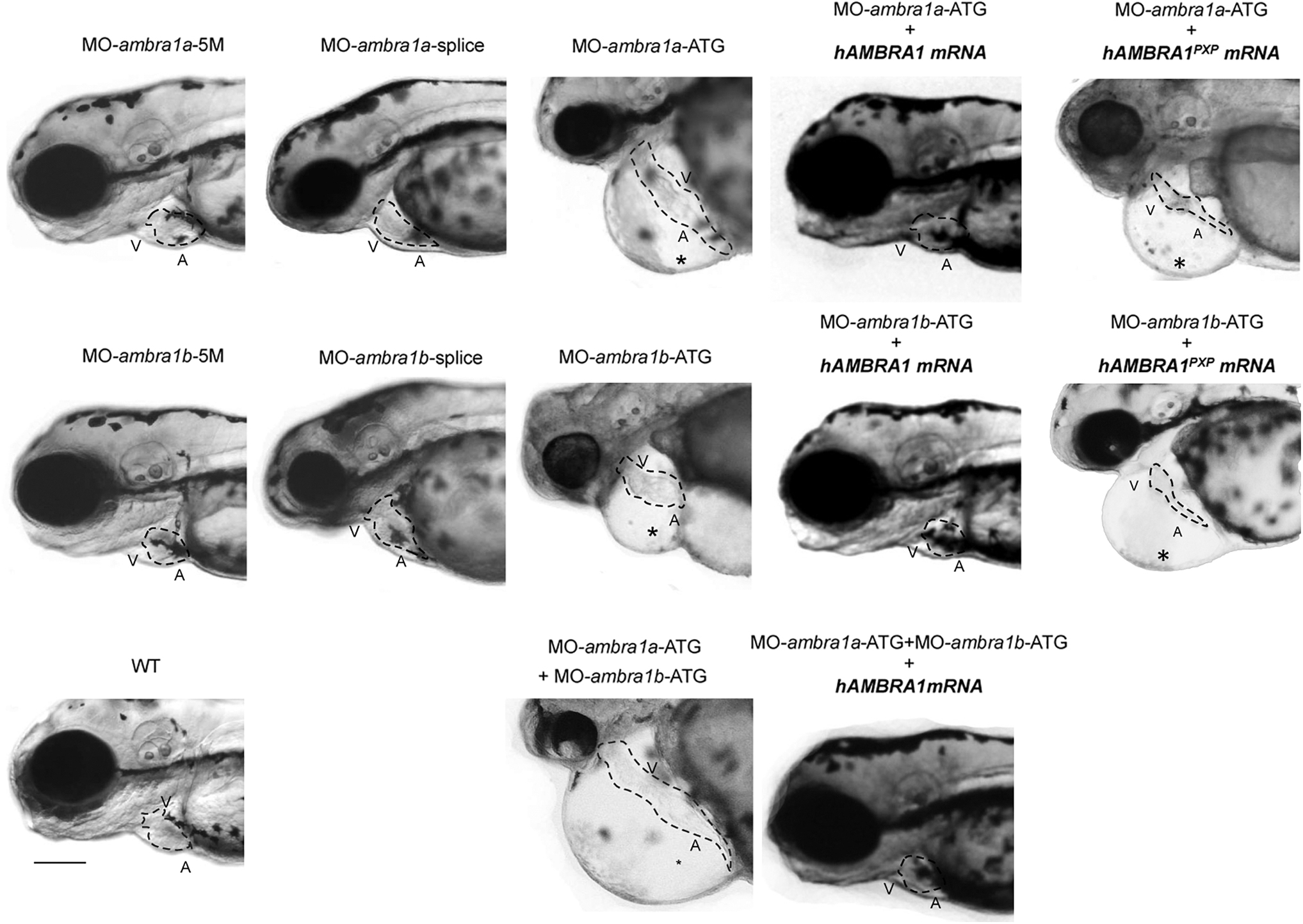
Fig. 1 Knockdown of zebrafish ambra1a and ambra1b impairs cardiac development. Representative bright-field images of the cardiac region of 3 dpf live embryos injected with the indicated MOs. ambra1a and ambra1b ATG-morphant embryos show pericardial edema (asterisk) and string-like heart tube when compared to WT and 5M control embryos. No visible abnormalities are evident in splice morphants, whereas cardiac defects are enhanced in the double morphants. The phenotypic defects of ATG-morphant embryos are rescued by co-injection with hAMBRA1 mRNA, but not with the AMBRA1PXP mRNA. The dotted line highlights the heart shape in the different conditions. A, atrium; V, ventricle. Scale bar, 200 μm. Images taken at 10 × magnification. More than 100 embryos observed for each condition, from five independent experiments. AMBRA1, activating molecule in beclin-1 (BECN1)-regulated autophagy; dpf, days postfertilization; hAMBRA1, human AMBRA1; MOs, morpholino oligonucleotides; mRNA, messenger RNA; WT, wild type.