
Fig. 3
|
Fig. 3
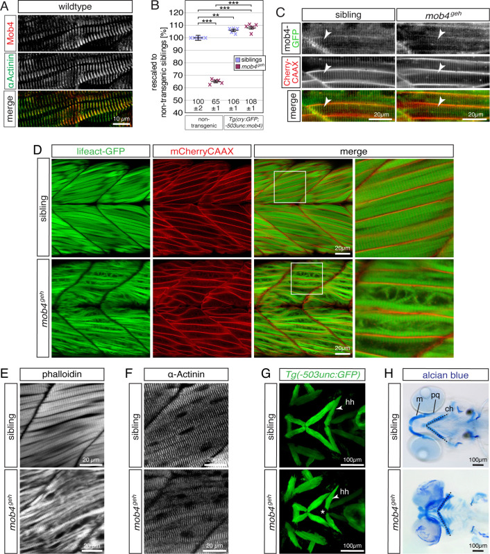
(A) At 3dpf, antibodies against human MOB4 colocalised with antibodies against the Z-disc protein α-Actinin (n = 6 per genotype). (B) At 3 dpf, the birefringence of mob4geh homozygotes was significantly higher in the transgenic background of Tg(cry:GFP;-503unc:mob4). Also compared to non-transgenic siblings (100 ± 2%), the birefringence of Tg(cry:GFP;-503unc:mob4) transgenic mob4geh homozygotes (108 ± 1%) and siblings (106 ± 1%) was significantly higher. Crosses represent averaged birefringence of clutches with a minimum of 4 larvae per genotype (n = 5 clutches). Data are presented as mean ± SEM; *** P < 0.001 and ** P < 0.01 by one-way ANOVA with post hoc Tukey’s test. (C) In 3-dpf-old siblings and mob4geh homozygotes, Mob4-GFP fusion protein (green) expressed via Tg(cry:GFP;-503unc:mob4-GFP) colocalised to t-tubules (red, arrowhead) marked in the Tg(acta1:mCherryCAAX) transgenic background (n = 3 per genotype). (D) Highlighting F-actin with transgenic Tg(acta1:lifeact-GFP) in green confirmed residual myofibril striation and revealed disorganised thin filaments within mob4geh homozygotes at 3 dpf. Sarcolemma and t- tubules were labelled by mCherry fluorescence (red) in the Tg(acta1:mCherryCAAX) transgenic background (n = 6 per genotype). Boxed areas are magnified. (E) Labelling of F-actin with phalloidin revealed that the robust myofibril striation of siblings was reduced in mob4geh homozygotes at 3 dpf (n = 6 per genotype). (F) At 3 dpf, antibodies against α-Actinin that mark sarcomere’s Z-disks showed the typical striation of the myofibril in siblings and mob4geh homozygotes (n = 4 per genotype). (G) Visualisation of the cephalic muscles in the transgenic Tg(−503unc:GFP) background revealed that, in contrast to siblings, a gap was formed between the two hyohyoideus (hh) muscles in mob4geh homozygotes at 3 dpf (representative Z-stacks) (n = 3 per genotype). (H) At 6 dpf, representative Z-stack projections of Alcian blue stained larvae depicted cartilage malformations in mob4geh homozygotes and a widened angle formed by the two ceratohyal cartilage structures (dotted lines) (n = 4 per genotype). Designations: ceratohyal (ch); Meckel’s cartilage (m); palatoquadrate (pq). Scale bar sizes are indicated.