
|
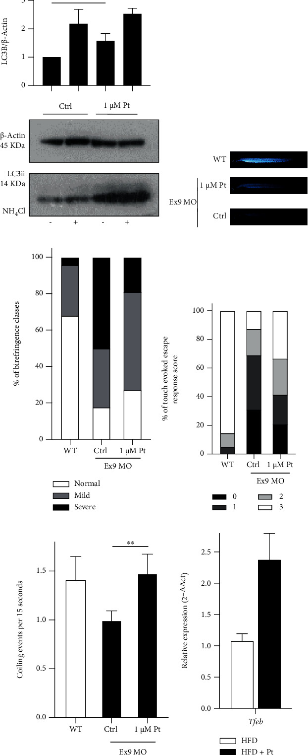
Figure 6 Pt-induced autophagy and TFEB activation in vivo. (a–e) Evidence of Pt-induced autophagy and functional improvement in dystrophic zebrafish morphants with altered Collagen VI. (a) Representative Western blot and quantification of the lipidated form of LC3 in zebrafish embryos treated as indicated. Mean values + SEM; N ≥ 3. (b, c) Birefringence assay. (b) Representative images from WT fish or ColVI morphants (Ex9 MO) after treatment with Pt or 0.1% DMSO (Ctrl). (c) 1 μM Pt induced a significant increase in the birefringence of morphants at 48 hpf. The percentage of fish showing a severe phenotype of ColVI-related myopathy was considerably reduced by 1 μM Pt (Pt vs Ctrl: p =0.016). Mean values + SEM. N ≥ 60 embryos for each condition, from at least 3 separate experiments. (d) Touch-evoked response assay. 1 μM Pt increased the percentage of fish showing mild or normal phenotypes (p = 0.0007). Responses were evoked by touching 48 hpf embryos with a tip. N ≥ 70 events for each condition, from at least 3 separate experiments. (e) Spontaneous coiling events of ColVI morphants measured at 24 hpf after 3 hours of treatment with Pt. 1 μM Pt completely rescues the deficit of spontaneous movements. Mean values + SEM. N ≥ 100 events for each condition, from at least 3 separate experiments. (f) RT-qPCR of Tfeb transcription in inguinal white adipose tissues from obese (HFD) and Pt-treated (HFD + Pt) mice. Mean values + SEM, N ≥ 14 mice for each condition.