
|
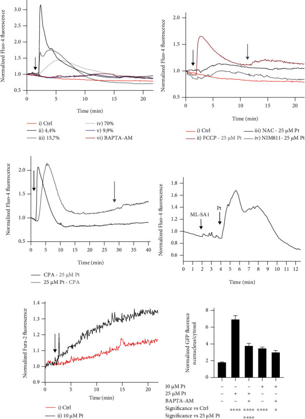
Figure 4 Pt elicits Ca2+ signaling in HeLa cells; N > 36 for each time point and condition, observed in at least 3 separate experiments. (a) Cytosolic Ca2+ signals induced by 25 μM Pt (Fluo-4 fluorescence). The arrow indicates the addition of 0.1% DMSO in curve i) and of 25 μM Pt in curves ii), iii), iv), v), and vi). The cells in curve vi) were pretreated for 20 min and continued to be incubated after Pt addition with 5 μM BAPTA-AM. (b) Cytosolic Ca2+ signals elicited by 25 μM Pt after pretreatment of the cells and in the continuing presence of modulators. 25 μM Pt was added when indicated by the black arrow for curves iii) and iv), when indicated by the grey arrow for curve ii). Modulators were as follows: ii) 2 μM FCCP, added when indicated by the black arrow (no preincubation); iii) 10 mM NAC (preincubation, 30 min); iv) 0.8 μM NIM811 (preincubation, 20 min); i) control: addition of DMSO (0.1% final concentration) when indicated by the black arrow. Please note the difference of the Y scale between panels (a) and (b). (c) Ca2+ mobilized by Pt largely originates from ER. Curve i): the SERCA inhibitor CPA (20 μM) was added when indicated by the black arrow; Pt (25 μM) when indicated by the grey arrow. Curve ii): the sequence of the additions was the opposite (Pt first). In both cases, excess EGTA was added to the medium 30 sec before the first addition. (d) The cellular Ca2+ mobilized by Pt in HeLa cells comes mostly from subcellular compartments other than the lysosomes (see text for details). Normalized Fluo-4 fluorescence. (e) Cytosolic Ca2+ signals elicited by 10 μM Pt measured with the ratiometric probe Fura-2. The arrow indicates the addition of: i) 0.1% DMSO; ii) 10 μM Pt. The difference between the two curves is significant ( p < 0.05 ) from t = 5 min onwards. (a–e) Error bars are omitted for clarity. (f) TFEB migration in HeLa cells exposed to the indicated conditions for 3 hours. Mean values + SEM.