IMAGE

Fig. 7

ID
ZDB-IMAGE-200730-15
Source
Figures for Hall et al., 2020
Image
Figure Caption

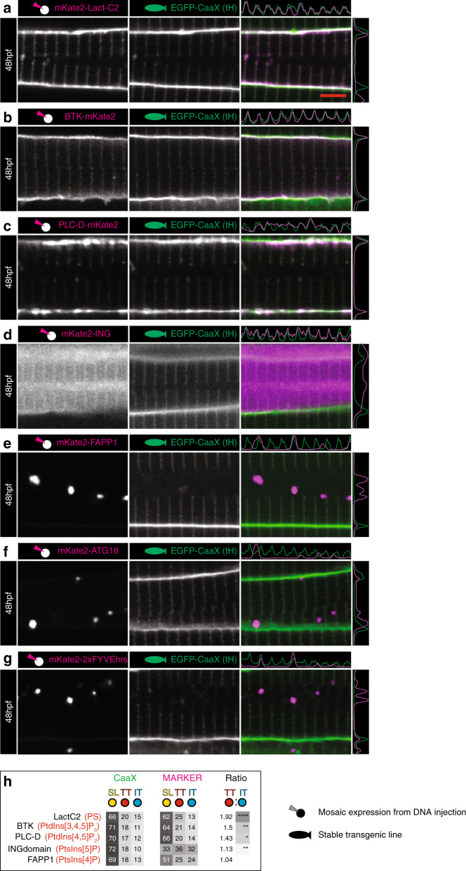
Fig. 7 Phosphoinositide signature of early T-tubules.

a LactC2 probe for phosphatidylserine (PS). b BTK probe for PtdIns(3,4,5)P2. c PLC-D probe for PtdIns(4,5)P2. d ING probe for PtdIns(5)P. e FAPP1 probe for PtdIns(4)P. f ATG18 probe for PtdIns (3,5)P2. g 2xFYVEhrs probe for PtdIns(3)P. h Ranked heatmap comparing average pixel intensity across each domain (T-tubule, sarcolemma and inter-T-tubule domain), for markers of interest and EGFP-CaaX. n = 6 fibres per marker, each from individual biologically independent animals per condition, measured over one independent experiment. Two-tailed T-test. *p < 0.05, **p < 0.01, ***p < 0.001, ****p < 0.0001. Exact p values are given in source data. Scale bars, 5 µm. All images are representative of 12 individual cells within different individual animals.

Acknowledgments
This image is the copyrighted work of the attributed author or publisher, and ZFIN has permission only to display this image to its users. Additional permissions should be obtained from the applicable author or publisher of the image. Full text @ Nat. Commun.