
|
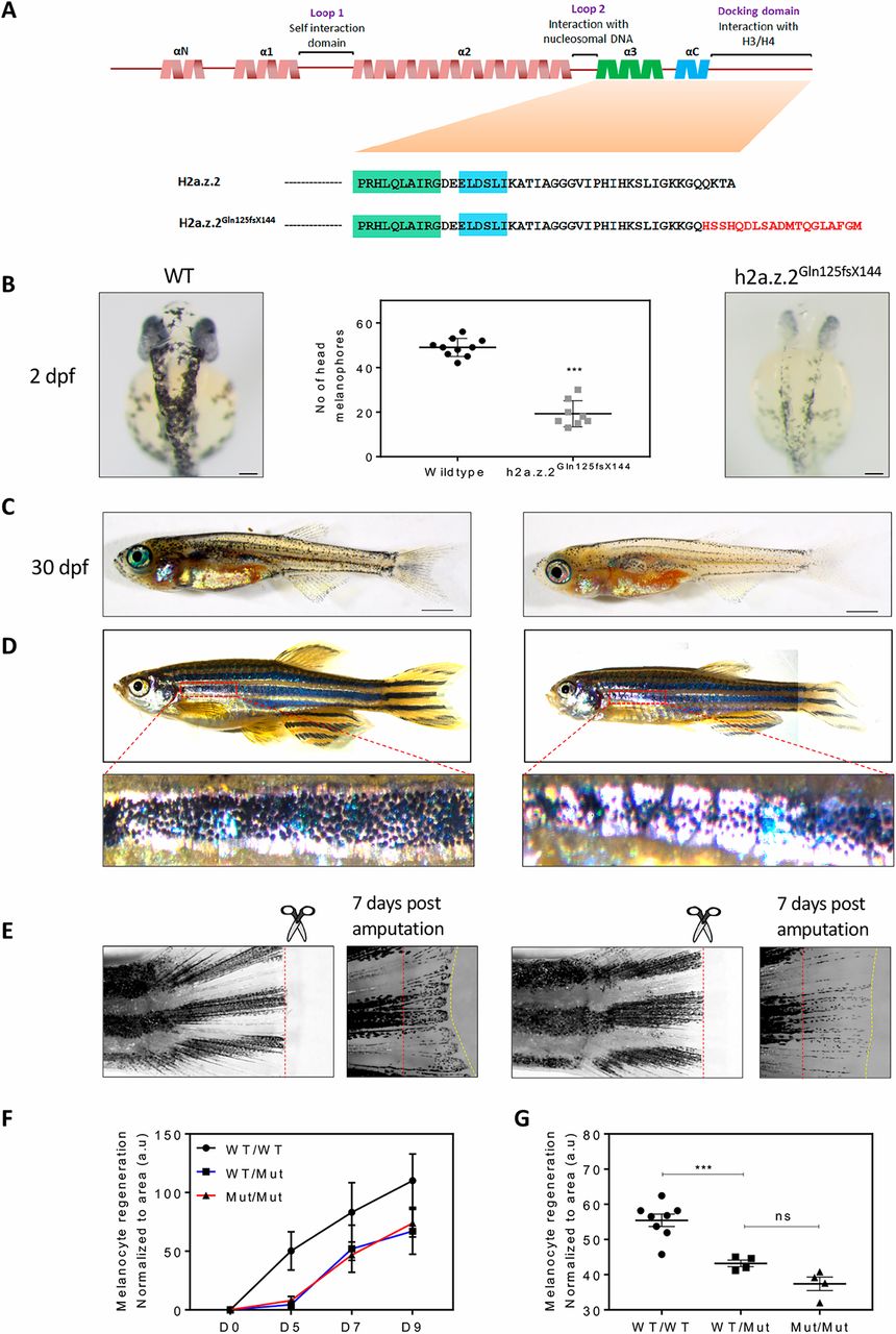
Fig. 6 Targeted mutation of h2a.z.2 affects melanocyte specification and regeneration. (A) Top: orthology-based schematic of the secondary structure of zebrafish H2AZ.2. The functional elements in the protein are denoted above. Below: amino acid sequence in H2A.Z.2 and the CRISPR mutant (H2a.z.2Gln125fsX144) generated in this study. (B) Representative images of wild-type and Z2 mutant embryos at 2 dpf. Right: wild-type sibling control. Left: homozygous H2a.z.2Gln125fsX144 mutant embryo. Middle: quantification of head melanophore numbers. Scale bar: 100 µm. (C) Representative images of wild-type and Z2 mutant juveniles at 30 dpf. Scale bars: 1 mm. (D) Representative images of wild-type and Z2 mutant adult animals. Higher magnification images of the first ventral stripe are shown below. (Adult fishes were imaged in parts and stitched.) (E) Fin clip and regeneration images of adult wild-type and Z2 mutant animals. Red-dashed lines indicate the site of amputation and the yellow-dashed line indicates the margin of the regenerated fin. (F) Kinetics of melanophore pigment regeneration over a period of 9 days after fin amputation (n=4). (G) Quantification of melanophore pigment across wild-type (WT), homozygous and heterozygous H2a.z.2Gln125fsX144 mutants upon regeneration.