IMAGE

Fig. 7

ID
ZDB-IMAGE-190723-341
Source
Figures for Veloso et al., 2019
Image
Figure Caption

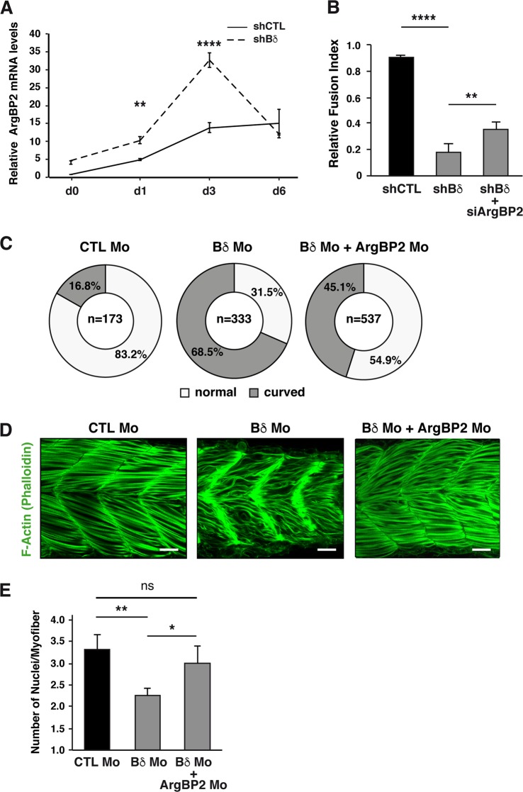
Fig. 7 PP2A-Bδ controls myoblast fusion through regulation of <italic>ArgBP2</italic> expression.

a RT-qPCR analysis of ArgBP2 mRNA levels in control (shCTL) and Bδ-KD (shBδ) C2C12 myoblasts during differentiation. Values are mean ± SD from three independent experiments. Two-way anova, with Bonferroni correction, ****P< 0.0001, **P< 0.01. b Fusion index of control (shCTL) and Bδ-knocked down (shBδ) C2C12 myoblasts transfected with a control siRNA or an siRNA against ArgBP2 (siArgBP2) from three independent experiments. One-way anova, with Tukey’s post-hoc test. **P< 0.01, ****P< 0.001. c–e Quantification of the curved trunk phenotype (c), d confocal imaging of F-actin (phalloidin, green, n = 10 for CTL Mo and n = 15 for Bδ Mo) and e quantification of the number of nuclei in fast skeletal myofibers of in 48-hpf zebrafish embryos injected with a control morpholino (CTL Mo, n = 5), or with a Bδ morpholino alone (Bδ Mo1, n = 3) or together with an ArgBP2 morpholino (Bδ Mo + ArgBP2 Mo, n = 10). Scale bars are 25 µm. One-way anova with Tuckey’s post-hoc test, **P< 0.01, *P< 0.05, ns: not significant

Figure Data
Acknowledgments
This image is the copyrighted work of the attributed author or publisher, and ZFIN has permission only to display this image to its users. Additional permissions should be obtained from the applicable author or publisher of the image. Full text @ Cell Death Dis.