
|
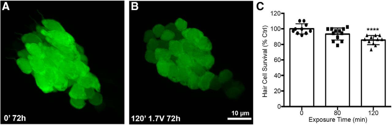
Fig. 4
Acoustic stimulation produces an exposure time-dependent reduction in saccular hair cells. Unexposed (A) and acoustically stimulated (B) saccules from myo6b:EGFP transgenic zebrafish. There was no obvious spatial pattern to the damage in the acoustically exposed saccules. C, Treatment with 1.7 V of acoustic stimulation for 120 min significantly reduces saccular hair cell number when assessed 72 h post-exposure (one-way ANOVA; exposure time: F(2,30) = 11.89, p = 0.0002). Asterisks indicate significant difference from unexposed age-matched control (****p < 0.001). N = 10–12 animals per treatment, values represent mean ± SD.