
|
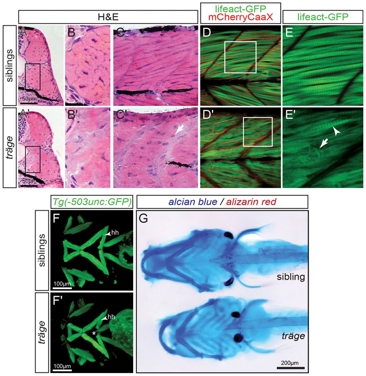
Fig. 3 Muscle phenotype of tmod4trg. (A) In comparison to their siblings, (A′) myofibres in H&E-stained cross sections of tmod4trg larvae show less defined cell shapes and inhomogeneous eosin staining at 3 dpf. (B,B′) Magnified views of the respective boxes indicated in A and A′. (C,C′) Accordingly, myofibres on sagittal sections of tmod4trg mutants appear unstructured, and blue haematoxylin-stained nuclei (arrow) are rounder in shape compared with those of siblings. (D) In a sagittal view of 3-dpf-old siblings, the myofibrils show typical striation, when marked by using the transgenic line Tg(acta1:lifeact-GFP). In addition, the transgenic background Tg(acta1:mCherryCAAX) labels the sarcolemma in red, showing that myofibrils occupy most of the myofibres. (D′) In matching tmod4trg mutants, the myofibril striation is rarely seen (arrowhead in E′), instead myofibres are filled with misorientated thin filaments (arrow in E′). (E,E′) Magnifications of respective boxes in D and D′. (F,F′) GFP highlights the cephalic musculature in the transgenic background of Tg(-503unc:GFP), here shown as ventral views of z-stack projections. (F) In contrast to their siblings, (F′) the two hyohyoideus (hh) muscles in tmod4trg mutants leave a gap between each other (asterisk). (G) Focus stacks of larvae that were stained with Alcian blue and Alizarin red depict cartilage malformations in tmod4trg at 6 dpf.