
|
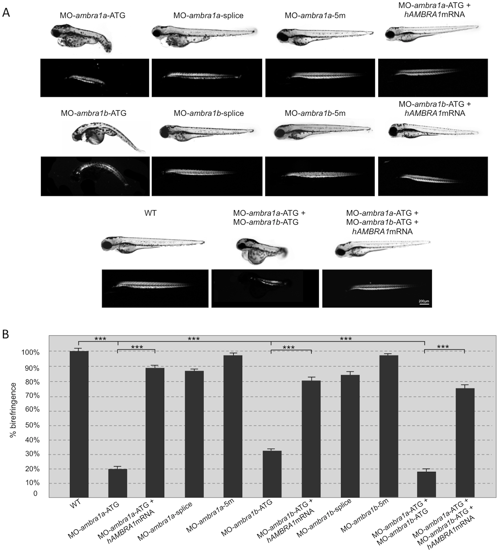
Fig. 1
Ablation of ambra1 results in reduced birefringence in zebrafish embryos.
(A). Representative images under normal and polarized light of 3-dpf live embryos injected with the indicated MOs. ATG-morphant embryos show reduced size, curved shape, pericardial edema and reduced birefringence when compared to WT and 5 m-control embryos. No visible abnormalities are evident in splice-morphants. The phenotypic defects of ATG-morphant embryos, including birefringence, are rescued by co-injection with 10 ng/embryo of human AMBRA1 mRNA. (B). Quantification of embryo trunk muscles birefringence shows a severe and statistically significant reduction in ambra1 ATG- and in co-injected morphants. The birefringence is faintly reduced in ambra1 splice-morphants, whereas WT and 5 m-control embryos display highly birefringent skeletal muscles. Muscle birefringence is statistically increased when ATG- and co-injected morphants are co-injected with human AMBRA1 mRNA (***, P<0.001).