
|
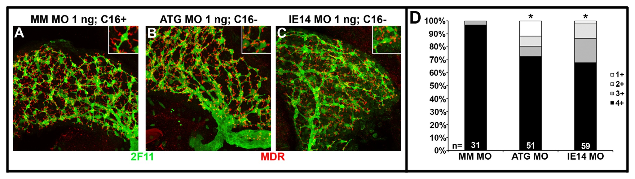
Fig. 5
Cirhin-deficient larvae have defects in biliary and canalicular morphology.
(A-C) Confocal projections of 5 dpf livers immunostained with antibodies against canalicular marker MDR (red) and biliary epitope 2F11 (green). Livers from fish with normal BODIPY-FL C16 processing (A) show elongated canaliculi intersecting with normally arborized biliary networks. In contrast, fish with reduced fluorescent lipid processing (B and C) show truncated, rounded canalicular profiles and more sparse biliary networks. Insets show high-magnification view of one or few cells to highlight canalicular morphology. (D) Semiquantitative scoring of canalicular morphology, with 1+=0-25% elongated, 2+=26-50% elongated, 3+=51-75% elongated, and 4+=76-100% elongated. *, p<0.05 vs Mismatch MO 1 ng.