
|
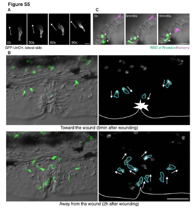
Fig. S5 Imaging uropod events (related to Figure 5)
(A) GFP-UtrCH (a biosensor for stable F-actin) labels the lateral sides of a neutrophil. Stable F-actin is localized at the lateral sides in addition to the tail during neutrophil random migration in the mesenchymal tissues of the head. The white arrows indicate direction of migration.
(B) GFP-UtrCH (a biosensor for stable F-actin) labels the tail and lateral sides of neutrophils when they migrate towards or away from a wound in the fin of Tg(MPO:EGFP-UtrCH)uw (movie S10).
(C) Inhibition of RhoA makes the tail round. EGFP-rGBD (RhoA binding domain of rhotekin fused to EGFP) was overexpressed into Tg(MPO:mCherry)uw to inhibit RhoA activity (Worthylake et al., 2001). Inhibition of RhoA activity disturbs cell migration in vivo and induces a rounded morphology of the tail (movie S12B). The arrow indicates the leading edge, which appears normal, while the arrowhead shows the round tail. Note that the magenta neutrophil expressing only mCherry migrates normally. See also movie S12C, in which RhoA is disturbed by overexpression of Rho T19N. Scale bars, 10 μm
(A), 50 μm (B), 20μm (C).
Reprinted from Developmental Cell, 18(2), Yoo, S.K., Deng, Q., Cavnar, P.J., Wu, Y.I., Hahn, K.M., and Huttenlocher, A., Differential Regulation of Protrusion and Polarity by PI(3)K during Neutrophil Motility in Live Zebrafish, 226-236, Copyright (2010) with permission from Elsevier. Full text @ Dev. Cell