
|
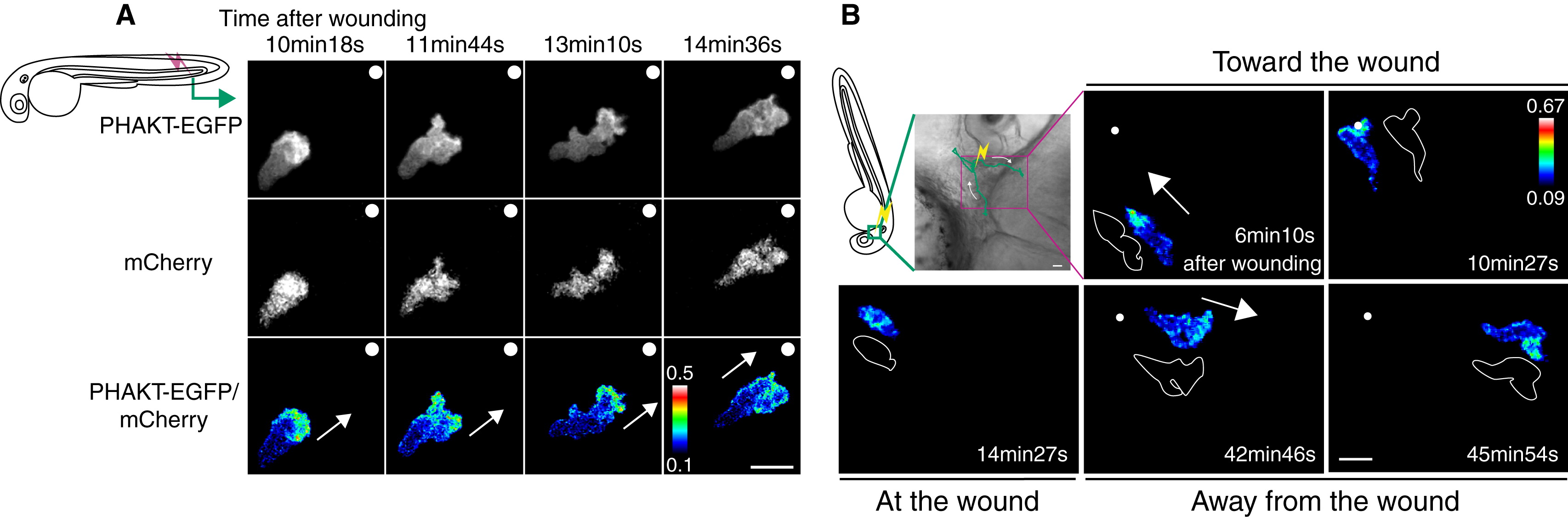
Fig. 2 PHAKT-EGFP Translocates to the Leading Edge When Neutrophils Come To and Leave Laser-Induced Wounds
(A) Time-lapse ratiometric imaging (PHAKT-EGFP/mCherry) reveals PI(3,4,5)P3-PI(3,4)P2 localization at the leading edge during attraction to a laser wound in the tail fin (>Movie S2A). The white dots and arrows indicate the position of the wound and direction of migration respectively. Scale bar = 10 μm.
(B) Reversal of PI(3,4,5)P3-PI(3,4)P2 (ratiometric imaging of PHAKT-EGFP/mCherry) when a neutrophil leaves the laser wound (Movie S2B). Note loss of PI(3,4,5)P3-PI(3,4)P2 polarity at the wound, followed by reversal of polarity to the opposite pole away from the wound when the neutrophil leaves the wound (green line, tracking of a neutrophil; yellow thunder, position of the laser wound; white arrows, direction of migration; illustration with white line, morphology of a neutrophil). Data are representative of more than five time-lapse movies from a minimum of three separate experiments. The numerical values of ratiometric analysis are shown in the scales. Scale bar = 10 μm.
Reprinted from Developmental Cell, 18(2), Yoo, S.K., Deng, Q., Cavnar, P.J., Wu, Y.I., Hahn, K.M., and Huttenlocher, A., Differential Regulation of Protrusion and Polarity by PI(3)K during Neutrophil Motility in Live Zebrafish, 226-236, Copyright (2010) with permission from Elsevier. Full text @ Dev. Cell