
|
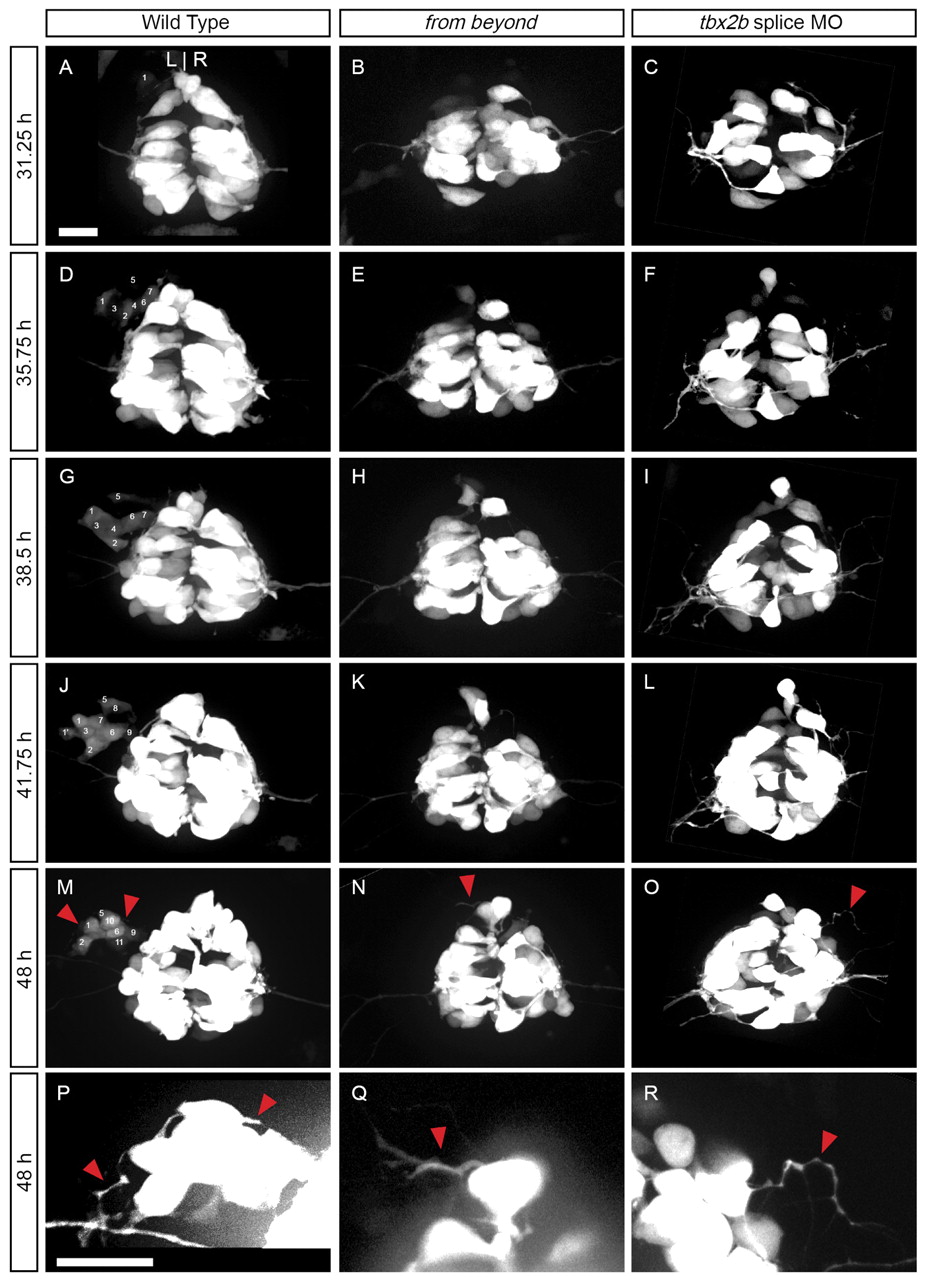
Fig. 8 Parapineal cells do not migrate to the left side of the brain in fby mutants. Dorsal views of foxd3:gfp expression in live (A,D,G,J,M,P) WT, (B,E,H,K,N,Q) fby mutant or (C,F,I,L,O,R) tbx2b splice morpholino-treated embryos. The data are snapshots of time-lapse confocal movies imaged from 31 to 48 hpf. P,Q,R are magnified and contrast-enhanced regions of the anterior pineal complex from M,N,O, respectively. In WT embryos, GFP-labeled cells emerged from the anterior left end of the pineal complex as a chain of cells between 31 and 39 hpf. Cells are numbered in the order of their appearance. Cell 1 divided from 39.75 to 40.25, and its daughter is labeled 1′. Note that some parapineal cells (e.g. #3, 4, 7, 8) become hidden by overlying cells. Between 39 and 48 hpf, the parapineal cells compacted together, and extended long thin projections (red arrowhead). By contrast, fby mutants and tbx2b morpholino-treated embryos had no GFP-labeled cells migrating to the left, but cells at the anterior end of the pineal complex still extended long thin projections. Scale bar: 25 μm.