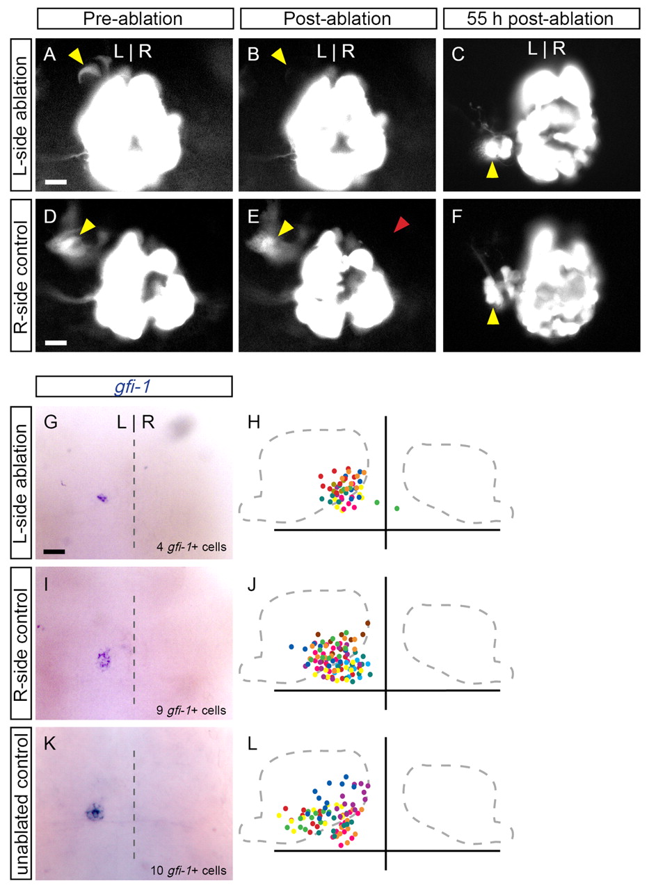
Fig. 5 Reduced numbers of parapineal cells are able to migrate to the left side of the brain. Dorsal views of the epithalamus in (A,B,D,E) 31-hpf zebrafish embryos or (C,F,G-L) 55-hpf larvae. Expression of foxd3:gfp labels parapineal precursor cells (A, yellow arrowhead), which are subsequently ablated with laser pulses (B). The remaining parapineal cells (average of 5±2 cells, n=18) migrate to the left side of the brain, as revealed by foxd3:gfp (C) and gfi1 (G) expression. In H, the position of gfi1-expressing cells in ten larvae are overlaid, with different colors representing individual samples. (D-F) As a control, cells contralateral to the parapineal precurors were ablated (red arrowhead). The number of parapineal cells (10±1, n=8) and their migration in these controls (I,J) were identical to those in unablated controls (K,L; 10±2 cells, n=10). Scale bars: 25 μm.
Image
Figure Caption
Acknowledgments
This image is the copyrighted work of the attributed author or publisher, and
ZFIN has permission only to display this image to its users.
Additional permissions should be obtained from the applicable author or publisher of the image.
Full text @ Development

