
|
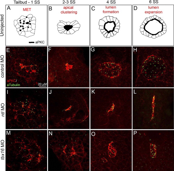
Fig. 7 Ntl and Tbx16 regulate distinct steps of KV development. Co-immunostaining of aPKC and aTubulin in KV cells (E–P) at four time-points defined temporally distinct steps of KV development in wild-type (represented as illustrations in panels A–D) and control MO injected embryos (E–H). These steps include a mesenchymal to epithelial transition (MET) marked by apical membrane biogenesis in KV cells (A, E), aggregation of apical membranes into a central cluster (B, F) and formation (C, G) and expansion (D, H) of the fluid-filled KV lumen. For simplification, cilia were excluded from the diagrams in panels A–D. In ntl morphant embryos, apical membranes were established (I) and clustered (J), but the KV lumen failed to inflate (K, L). Apical foci were present in tbx16 morphants (M), but KV cells did not aggregate (N). Lumen formation proceeded without apical clustering, giving rise to dysmorphic and/or multiple vesicles (O, P). All confocal micrographs are at the same scale (see scale bar in panel E).
Reprinted from Developmental Biology, 310(2), Amack, J.D., Wang, X., and Yost, H.J., Two T-box genes play independent and cooperative roles to regulate morphogenesis of ciliated Kupffer's vesicle in zebrafish, 196-210, Copyright (2007) with permission from Elsevier. Full text @ Dev. Biol.