
Fig. 4
|
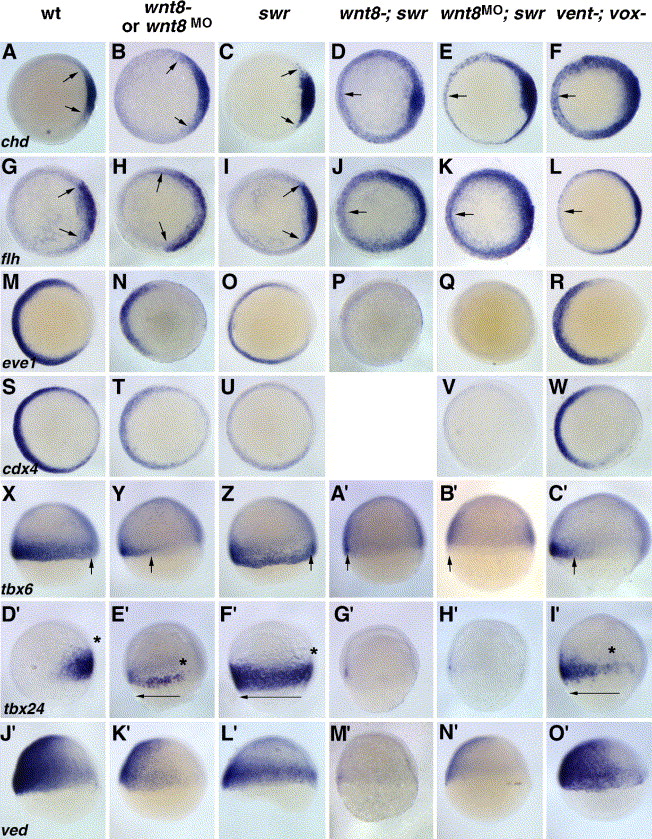
Fig. 4 Both wnt8; swr and vent; vox double mutants display expansion of axial mesoderm, but only wnt8-; swr embryos show loss of non-axial mesoderm. In situ hybridizations for chd (A–F), flh (G–L), eve1 (M–R), cdx4 (S–W), tbx6 X–C′), tbx24 (D′-I′), and ved (J2-O′). Genotypes are indicated above each column. All embryos are at shield stages except panels X–C′ (60% epiboly) and D′–I′ (70% epiboly). Both wnt8; swr (D, J) and vent; vox (F, L) double mutants show a loss of axial mesoderm repression, although flh expansion is less severe in vent; vox mutants than in wnt8; swr mutants (arrow in panel L, compare to panel J). All non-axial–mesoderm markers (eve1, cdx4, tbx6, tbx24, and ved) are strongly reduced or absent in wnt8; swr double mutants (P, V, A′, G′, M′) but are still expressed in vent; vox mutants (R, W, C′, I′, O′). Asterisks in panels D′-I′ represent the dorsal limit of tbx24 staining. (A–W) Animal views, dorsal right. (X–O′) Lateral views, dorsal right, anterior up.
Reprinted from Developmental Biology, 287(2), Ramel, M.C., Buckles, G.R., Baker, K.D., and Lekven, A.C., WNT8 and BMP2B co-regulate non-axial mesoderm patterning during zebrafish gastrulation, 237-248, Copyright (2005) with permission from Elsevier. Full text @ Dev. Biol.