
Fig. 1
- ID
- ZDB-FIG-170428-13
- Publication
- Klangnurak et al., 2017 - Fine selection of up-regulated genes during ovulation by in vivo induction of oocyte maturation and ovulation in zebrafish.
- Other Figures
- All Figure Page
- Back to All Figure Page
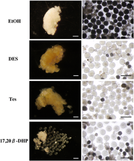
The in vivo bioassay was performed at a final concentration of 5 μM of DES, 1 μM of Tes or 0.01 μM of 17, 20β-DHP. One side of the ovary was observed by stereomicroscopy. The morphologies of the ovarian samples after three hours of treatment by EtOH, DES, Tes and 17, 20β-DHP were photographed. Ovaries before (left panels) and after (right panels) splitting are shown. After treatment with EtOH, the oocytes remained opaque and showed no morphological change after exposure to water. Oocytes after treatment with DES or Tes became transparent. A fertilization membrane developed in oocytes ovulated by 17, 20β-DHP treatment after exposure to water. Scale bars indicate 1 cm |