|
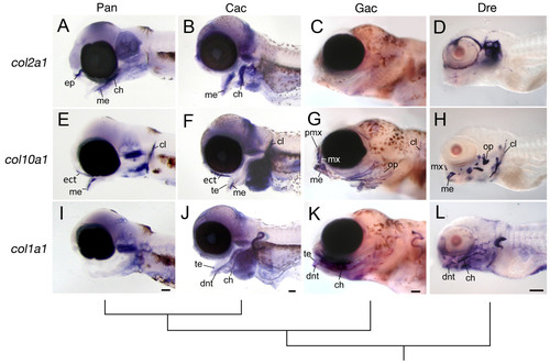
Whole-mount in situ hybridization for collagen gene expression in the pharyngeal skeleton. Lateral views are shown of embryos for P. antarcticum, C. aceratus, G. aculeatus, and D. rerio at comparable stages of development. (A, E, I) P. antarcticum, mid-larvae; (B, F, J) C. aceratus, mid-larvae; (C, G, K)G. aculeatus, 11 dpf; and (D, H, L) D. rerio, 5 dpf. Notothenioids retain strong col2a1 expression in the pharyngeal skeleton (A, B) well into larval development compared to both G. aculeatus (C) and D. rerio (D). Conversely, col10a1 (E, F) and col1a1 (I, J) expression is relatively weak or absent in the notothenioids. The tree represents the evolutionary relationship among species. Abbreviations: ch, ceratohyal; cl, cleithrum; dnt, dentary; ep, ethmoid plate; ect, ectopterygoid; me, Meckel′s cartilage; mx, maxilla; op, opercle; pmx, premaxilla; te, teeth. Scale bars, 100 μm.
|